Michael Hooker Microscopy Facility (MHMF.ORG)
![]()
|
|
Michael Hooker Microscopy Facility (MHMF.ORG) |
|
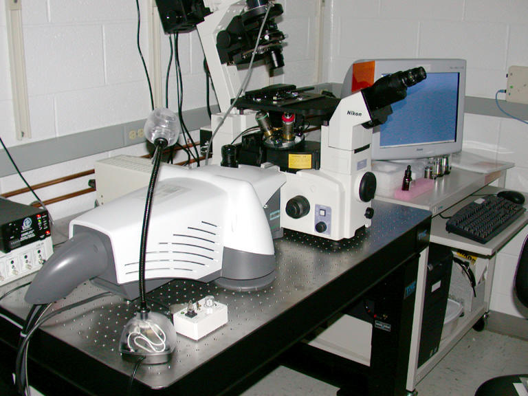
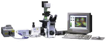
Perkin Elmer Yokogawa Nipkow disk confocal
Introduction:
This confocal system has a Yokogawa multiple lens Nipkow spinning disk scanner (CSU10), allowing it to capture confocal images rapidly with a minimum of excitation light. Images are captured using a Hamamatsu OrcaER camera driven by the SimpelPCI software from Hamamatsu. Excitation light is provided by a ~100 mW Krypton/Argon laser, which can excite at 488, 568 & 647 nm. Imaging of FITC-like, Rhodamine-like, Cy5-like and similar fluorophores, including GFP, Calcium Green & Texas Red can be carried out. A filter wheel changes emission filters. A Prior motorized drive is attached to the fine focus knob for software control of z- position. Transmitted light shutter permits widefield, DIC or phase contrast images to be interlaced with fluorescent image capture. Acquisition modes can be combinations of z-series, time lapse, &/or multiple fluorophores.
The confocal head is mounted on a Nikon TE2000E inverted microscope. Analysis software includes the Volocity 3D rendering & quantitation package & C-Imaging (including deconvolution on the \\Nomarski image processing workstation). Images can be transferred directly over the network.
Notes:
Operating the System
Equipment:
Objectives (These are the objectives shipping with the confocal. Other objectives will be available) Mag. NA type WD corrections cover slip Immersion part no. dic/phase condensor Wollaston 20x 0.75 S-Fluor 1.0 mm 0.17 (#1.5) air 93127 40x 1.3 S-Fluor 0.22 mm 0.17 (#1.5) oil 93130 100x 0.7-1.3mm S-Fluor 0.2 mm NA 0.17 (#1.5) oil
Filters
Position # Label Fluorophore Filterset Specifics 1 ---- Trans - - 2 UV DAPI Nikon UV-1A 3 B FITC, GFP Nikon B-2E/C Specifics 4 G Texas Red 96322 TR C109382 5 CY5 96324M CY5 HQ 6 not installed - CFP 96341 HQ: CY GFP C77389 ex=D436/20 65555, em=HQ480/40 66403 not installed - YFP 96345 HQ: Y GFP C74135 ex=HQ500/20 60341, em=HQ535/30 Chroma 41028
Position # Label Fluorophore Filterset Specifics 1 B FITC, GFP Nikon B-2E/C Specifics 2 G Rhodamine Nikon G-2E/C Specifics 3 4 5 6
| Position Ex Em |
| 1 488/10 525/50 |
| 2 568/10 600/45 |
| 3 PARK PARK |
| 4 647/10 700/75 |
| Triple Dichromatic mirror specifications |
Related Links:
|
|
|
Copyright 2001-2015 Dr. M. Chua, School of Medicine, University of North Carolina, Chapel Hill, NC 27599 |
| Go Back | Booking Resources |
Questions/comments/problems: Michael Chua |
|
|
Last Updated: 2014-07-24 |